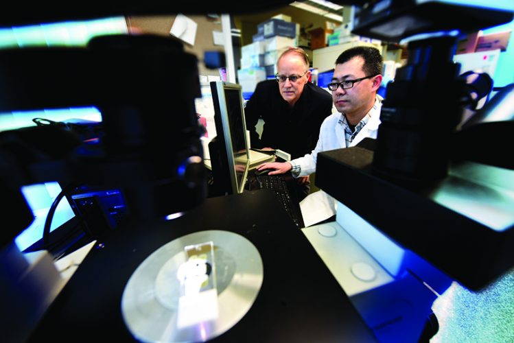
While we may joke about the differences between the brains of males and females, scientists like Dr. Darrell Brann are providing more and more evidence that one thing they have in common is estrogen.
It started with a song
While estrogen being made by the brain, never mind male brain, may seem a little counterintuitive, there is decades-old evidence that the widely known female sex hormone, has important roles in both sexes, including brain-related ones like protecting younger women from stroke and giving a male songbird his tune.
There is also the biophysical twist that estrogen comes from testosterone with the help of an enzyme called aromatase.
“You have to have testosterone to have estrogen,” says Dr. Lawrence C. Layman, chief of the MCG Section of Reproductive Endocrinology, Infertility and Genetics. While aromatase is the magic that makes the conversion, it’s estrogen – not aromatase or testosterone – that has the final say, Layman notes.
Masculinization of the brain is another great example. During the first days of life it’s estrogen that actually masculinizes the brain.
Transgender research, for example, has shown that mice exposed to estrogen shortly after birth, often in their bellies, exhibit male behavior, mounting females even if they are female, Layman says. When you block the estrogen effect, the mice act more like females. “So a male who feels he is really a female likely has less estrogen,” says Layman.
To complicate this scenario a bit more, the estrogen that masculinizes the brain starts with testosterone from the testes that gets converted to estrogen by aromatase in the hypothalamus, an area in the front of the brain with a lot of roles, including controlling emotional activity, sleep and growth.
But it’s still estrogen that prompts the masculinization, including the size, of different brain areas and resulting sexual behavior, says Brann, interim chair of the Department of Neuroscience and Regenerative Medicine at the Medical College of Georgia.
While doing his research fellowship at MCG in the late 1980s, Layman, a reproductive endocrinologist, had learned what most of us probably suspected but we have since learned isn’t true: that testosterone masculinized the brain and estrogen feminized it.
Still what scientists like Brann are realizing today is not that much of a stretch, says Layman.
Levels of systemic estrogen have long been tied to memory, with women reporting memory loss after menopause. Estrogen’s most established role of regulating reproductive function also actually happens from the brain, notes Layman.
In fact, it was in reproduction that Brann got his estrogen start.
A stroke of estrogen
Brann and his team showed nearly two decades ago that the neurotransmitter glutamate has an important role in regulating the hormones the brain releases to control ovulation. Glutamate was already a suspect in age-related memory problems, and Brann was beginning to dissect how it might impact the decreased fertility that also occurs with age. He was also growing an interest in what was happening to memory.
Within a few years, acknowledging the stroke and cardiovascular protection estrogen appears to give younger women, he also was exploring whether estrogen and estrogen-like drugs could help traumatized cells that encircle the core of a stroke, called the penumbra, survive and even encourage stem cells in the brain to mature into brain cells.
By 2008, Brann and his mentor Dr. Virendra B. Mahesh, chair emeritus of the MCG Department of Physiology and Endocrinology (now the Department of Physiology) had coedited a special issue of the journal Molecular and Cellular Endocrinology, that took on the emerging topic of estrogen’s action in the brain.
In fact, by then Brann, who completed his PhD in endocrinology at MCG with Mahesh, had shifted his research focus from estrogen’s impact on reproduction to its role in stroke recovery and the brain.
“There is no doubt reproduction is a primary function of estrogen, but estrogen has lots of functions, probably hundreds,” he said at that time.
Again, scientists have had evidence since the 1970s that aromatase has a presence in the brain and now knew that in both males and females, neurons and their supporting brain cells called astrocytes produced estrogen via aromatase made in the hippocampus and cerebral cortex in a variety of species, including humans. In fact, work in the male songbird in the early 1990s at the University of California, Los Angeles indicated that brain-derived aromatase and estrogen have an essential role in the bird’s ability to make a song. But the tools to actually study nonreproductive functions were just coming around.
Timing really is everything
Brann and international colleagues from North China Coal Medical University and the University of Texas Health Sciences Center at San Antonio reported in 2009 that estrogen therapy must be given soon after menopause – surgical or natural – to again enable stroke protection.
It was definitely a timely finding. The Women’s Health Initiative, which continues today in its second extension, was reporting some controversy about widely used estrogen therapy. The study, which followed more than 160,000 women ages 50-79, was finding that rather than providing protection, estrogen therapy actually increased risks for things like stroke and blood clots, heart disease, dementia and breast cancer.
Part of the controversy was that some of the women had a wide gap in between the time they hit menopause and started estrogen therapy and some, including Brann, had evidence that if it were instead a continuum, the benefits could hold.
Brann and others reported in The Journal of Neuroscience, for example, that with a significant gap, as much as 50% of estrogen receptors in key regions like the learning and memory center of the brain vanished. Brann’s follow up studies published in 2011 showed that when estrogen therapy was started shortly after hormone levels dropped, more estrogen receptors on the brain cells of aging rats survived. Follow up work in women, has indicated a five to 10 year timeframe as well to continue to reap positive cardiovascular benefits.
Two years later, in the journal Brain, Brann published more animal evidence of the brain benefit of estrogen, showing that when ovaries are removed before the usual age of menopause, it left the brain more vulnerable to stroke and Alzheimer’s. Estrogen therapy started right after the oophorectomy reduced the vulnerability.
Connecting the sex hormone
Fast forward two more years and Brann and his team received another $1.8 million grant from the National Institutes of Health to explore emerging evidence that aromatase, the enzyme that converts testosterone to estrogen, had a big role in both a healthy brain as well as an injured state like stroke or traumatic brain injury.
The MCG team worked with Dr. Ratna K. Vadlamudi, molecular biologist, professor of obstetrics and gynecology at the University of Texas Health Sciences Center and an expert in human nuclear receptors, including the estrogen receptor, to develop animal models that would enable the studies. They included a mouse with aromatase removed from neurons, another with it removed from astrocytes, and a third with it missing both places. The spectrum enabled the early exploration of the exact role of aromatase in each brain cell type. What they found was that with injury, aromatase and estrogen expression shifts more to the astrocytes, apparently to help these already supportive brain cells work even harder to help their neurons.
Estrogen fundamental to making memories
Just this year, again in The Journal of Neuroscience, Brann and Vadlamudi reported in both males and females, even more connections between estrogen and a healthy brain.
They found that when neurons don’t make estrogen, resulting mouse brains have significantly less dense spines and synapses – both key communication points for neurons – in the biggest part of their brain, called the forebrain.
“I think this gives us a new appreciation and understanding of how the brain forms memories, and that estrogen is important in that process,” Brann says of the work that scored in the top 5% of all research articles at the moment scored by Altmetric, a service that keeps tabs on who is paying attention to research.
Mice whose neurons didn’t make estrogen had impaired spatial reference memory – like a baseball player not knowing where home plate is and what it means to get there – as well as recognition memory and contextual fear memory – so they have trouble remembering what’s hazardous.
Restoring estrogen levels to the brain area rescued the impairments.
It was known that when aromatase is blocked, it can result in memory problems for animals and humans alike. In fact, patients who take an aromatase inhibitor for estrogen-dependent breast cancer have reported memory problems.
So for these studies they looked at mice where aromatase was knocked out of the forebrain, including the hippocampus, which has a role in making long-term memories and spatial memory, and the cerebral cortex, which is important to memory, attention, awareness and thought.
They depleted aromatase only in the excitatory neurons – called excitatory because they help make some action like a thought happen – in the forebrain as a way to focus on the role of estrogen produced by these brain cells.
The bottom line was a 70-80% decrease in aromatase and estrogen levels in the neurons in these areas of the brain. The other bottom line: “The knockout mice can’t remember as well as the normal mice,” Brann says.
They put male and female mice through extensive behavioral testing, including those that had their ovaries removed as a control.
A slice of brain with estrogen on top
Electrophysiological studies of slices of the estrogen-altered brain showed that while long-term potentiation – which is the process by which synapses strengthen to form a memory – worked, it didn’t function to nearly the same degree. But, putting an equivalent estrogen directly onto slices of the hippocampus restored that ability within minutes.
Interestingly, while memory deficits clearly occurred in both sexes when estrogen production was impaired, deficits were more significant in the females.
Brann considers estrogen’s profound effect on long-term potentiation the most interesting of the newest findings.
“Neurons actually make and use estrogen for their communication and for the establishment of memories,” he says. “To me that is the most fascinating thing.”
Knocking out aromatase also decreased expression of CREB, a major transcription factor known to play a key role in learning and memory, as well as neuron-nourishing brain derived neurotrophic factor, or BDNF.
A novel neuromodulator
The scientists say these findings implicate neuron-derived estrogen as a novel neuromodulator, basically a critical messenger one neuron relies on to communicate with others, which is essential to key functions like cognition.
“It’s direct genetic evidence of this role and I think that is important to have,” Brann says.
Neuromodulators need to be created and released quickly, Brann notes, which is how estrogen gets produced in the brain. We all have basal levels that can be rapidly induced when needed, he adds.
He theorizes that glutamate, the brain’s most abundant excitatory neurotransmitter and essential to learning and memory, prompts the neurons’ production of estrogen by inducing aromatase, Brann says. He notes that like many things in our body, glutamate is a double-edged sword that can also kill neurons, and that the brain has receptors to both use and eliminate the brain chemical.
Meanwhile they learned that astrocytes only seem to make estrogen in response to injury. In this apparent maneuver to step up support for neurons, it’s likely cytokines, substances secreted by immune cells to also make something happen, that prompt supportive astrocytes to start estrogen production. The scientists already have another project looking specifically at these supportive brain cells.
There are many blanks left to fill in before the tightly regulated system is understood well enough to be manipulated to help patients. “We are putting all the pieces together right now,” says Brann, who is excited and hopeful about future patient possibilities.
His future includes learning more about what is regulating brain aromatase and he’s thinking the answer may be glutamate. Also, whether brain estrogen and aromatase levels decrease with normal aging as he suspects and, if they do, what compounds might be used to increase aromatase and estrogen production in the brain, Brann says.
“We have some aged estrogen knockout animals now that we need to look at,” he says.
The ovaries also use aromatase, he notes, although a slightly different form, to convert testosterone to estrogen. Evidence to date, including the new study, indicates that removing the ovaries does not impact brain levels of estrogen, suggesting that one is not dependent on the other, Brann says. He notes that the estrogen made by the brain and ovaries is indistinguishable.
Brann also is now further exploring the way higher systemic estrogen levels in younger women also manage to reach the brain and perhaps afford that well-known extra protection premenopausal females exhibit against brain problems like stroke and Alzheimer’s.
He admits that when he started his studies years back, like so many others, he figured the circulating estrogen was the only estrogen helping the brain. When you took the ovaries out, the protection was gone, he says of countless animal studies, and parallel human evidence in women who had oophorectomies.
Today he and Layman concur that systemic levels of the hormone likely do provide women’s brain a bit of an edge. In fact, Brann has included mice with their ovaries removed in some of his studies to better assess the impact.
But it’s the estrogen in our brains that has his attention now.


