PROTEIN COCKTAIL
Patients with type 1 diabetes have significantly lower blood levels of four proteins that help protect their tissue from attack by their immune system, scientists report.
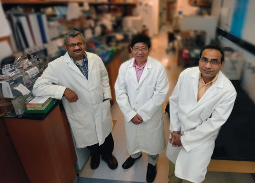
Conversely, their first-degree relatives, who share some of the high-risk genes but do not have the disease, have high levels of these proteins circulating in their blood, said Dr. Jin-Xiong She, director of the MCG Center for Biotechnology and Genomic Medicine.
Healthy individuals without the risky genes also have higher levels of the four proteins: IL8, IL-1Ra, MCP-1 and MIP-1ß, according to the study in the Journal of Clinical Endocrinology & Metabolism.
The scientists looked at 13 cytokines and chemokines, which are cell signaling molecules involved in regulating the immune response. They first looked at blood samples from children with type 1 diabetes and from individuals without antibodies to insulin-producing cells, a hallmark of this autoimmune disease. They then analyzed the blood of a second and larger set of individuals, which included 1,553 children with type 1 diabetes and 1,493 individuals without any sign of antibodies.
The findings point toward a sort of protein cocktail that could help at-risk children avoid disease as well as new blood biomarkers to aid in disease diagnosis, prognosis and management.
In this largest study of its kind, they consistently found that a higher percentage of type 1 diabetes patients had significantly lower levels of the same four proteins.
The research is funded by the National Institutes of Health and the Juvenile Diabetes Research Foundation.
SCHIZOPHRENIA FINDINGS

High blood levels of a growth factor for blood vessel development and brain cell protection correlate with an abnormally small size of brain areas key to complex thought, emotion and behavior in patients with schizophrenia, researchers report in the journal Molecular Psychiatry.
Higher blood levels of vascular endothelial growth factor, or VEGF, also correlate with high blood levels of interleukin 6, an inflammation-promoting cytokine that can cross the blood-brain barrier, said Dr. Anilkumar Pillai, neuroscientist in the Department of Psychiatry and Health Behavior. Inflammation is increasingly associated with schizophrenia, and high blood levels of IL-6 already have been found in these patients.
The findings not only shed light on the disease, but suggest that a blood test could replace expensive brain-imaging studies to confirm the diagnosis. “We are talking about a molecule where you can just draw blood and look at the lab profile,” said Pillai, the study’s corresponding author.
His lab earlier showed low brain levels of VEGF in schizophrenia patients, which could help explain their lower blood flow and brain volumes. “Decreased blood flow leads to decreased brain tissue volume,” he said. Inflammation also can reduce brain size.
STARRING ROLE
A star-shaped brain cell called an astrocyte appears to help regulate blood pressure and blood flow inside the brain, scientists report.
The finger-like appendages of astrocytes, called endfeet, wrap around the brain’s blood vessels, constantly monitoring the environment, said Dr. Jessica A. Filosa, neurovascular physiologist in the Department of Physiology. Findings indicate that when they sense a change in blood pressure, they release signals that help dilate or constrict the blood vessels, whichever it takes to maintain stasis.
“This is the first evidence of the astrocyte’s role in pressure-induced myogenic (muscle) tone, which is keeping things regular,” said Filosa, corresponding author of the study in The Journal of Neuroscience.
In fact, astrocytes keep their fingers on the pulse of blood vessels and neurons simultaneously, apparently playing an important role in balancing their needs. “They are perfect bridges between what is going on with neuronal activity and blood flow changes to the brain,” Filosa said.
Next steps include determining how the activation of astrocytes affects neuronal activity, so Dr. Ki Jung Kim, postdoctoral fellow and first author on the current study, is now recording from neurons as they change pressure inside blood vessels. The research was supported by the National Institutes of Health and the American Heart Association.
VITAMIN E BOOST
MCG scientists are drilling down on how vitamin E helps strengthen muscles.
They have shown that vitamin E, long known as a powerful antioxidant, is also vital in healing the protective outer covering of plasma.
“Every cell in your body has a plasma membrane, and every membrane can be torn,” said Dr. Paul L. McNeil, an MCG cell biologist and corresponding author of the study published in the journal Free Radical Biology and Medicine.
He and his colleagues administered a dye that cannot permeate an intact plasma membrane and found it easily penetrated the muscle cells of vitamin E-deficient rats. The finding has implications for conditions including muscular dystrophy, diabetes-related muscle weakness and traumatic brain injury.
“Part of how we build muscle is a natural tearing-and-repair process, and if that repair doesn’t occur, you get muscle cell death. If that occurs over a long period of time, you get muscle-wasting disease,” said McNeil. “[Our finding] means we know what the cellular function of vitamin E is, and knowing that cellular function, we can ask whether we can apply that knowledge to medically relevant areas.”
BRAIN AID
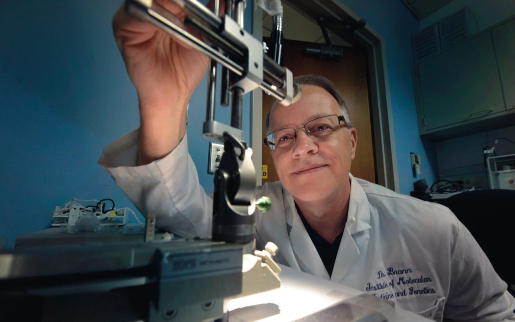
An enzyme that converts testosterone to estrogen appears to have significant impact in a healthy and injured brain, scientists report.
Evidence suggests that, in the brain, aromatase and the estrogen it enables neurons to produce helps keep brains healthy. Now, scientists are learning that with injury, aromatase and estrogen expression seem to shift to brain cells called astrocytes, helping support now-stressed neurons, said Dr. Darrell Brann, Regents’ Professor and vice chairman of the Department of Neuroscience and Regenerative Medicine.
Several studies, including those in Brann’s lab, have shown this primary shift in aromatase/estrogen expression from neurons to astrocytes following injury. In Brann’s case, the studies have been in the hippocampus, a center of learning, memory and emotions. When he used a drug to reduce astrocyte’s aromatase expression in that region, increased inflammation and brain damage resulted.
A new $1.8 million grant from the National Institutes of Health will enable him to further elucidate the role of aromatase and estrogen in injury and in health and, ideally, point toward therapies that can augment the brain’s apparent effort to heal.
MUSCLE MIGHT
Even without losing fat, more muscle appears to go a long way in fighting off the bad cardiovascular effects of obesity – a finding that could open the door to new treatments for cardiovascular disease.
“We understand that if you exercise, things get better,” said Dr. David Stepp, an MCG vascularbiologist. “What we don’t really understand is what about exercise is good.”
Stepp and his colleagues have evidence that muscle mass plays a significant role. While fat stores fuel, “muscle is a much more metabolically active tissue,” Stepp said. “It burns more oxygen at rest, it burns more energy at rest, so it burns more calories at rest.”
His team’s $2.2 million grant from the National Heart, Lung and Blood Institute is helping them determine if muscles secrete something that improves glucose metabolism. “We are trying to establish links between the health of skeletal muscles and the circulatory system,” said Dr. David Fulton, director of the MCG Vascular Biology Center and co-principal Investigator with Stepp on the grant. “When you are young, most of your body mass is skeletal muscle, so that glucose is efficiently distributed in the places where it should go to get used for energy and work.”
Stepp and Fulton authored a 2014 study in the Journal of the American Heart Association that showed the benefits of adding muscle to obese bodies. When they deleted myostatin from mice – some normal and some genetically altered to be obese – both groups developed bigger muscles, and the normal mice had less fat tissue. Myostatin is a natural negative regulator of muscle growth.
But it was the obese-with-muscles mice that truly benefited: glucose tolerance and blood vessel dilation increased, and insulin resistance and superoxide production decreased. When they looked further at nonmuscular obese mice, they also found the superoxide-producing gene Nox1 is a major culprit in obesity-related vascular disease.
Obese mice with no muscle added but Nox1 removed also experience cardiovascular improvement, an observation that Dr. Jennifer Thompson, a GRU postdoctoral fellow, is pursuing further with an American Heart Association grant. Meanwhile, Stepp and Fulton are exploring how elevated blood glucose elevates Nox1, hoping to identify additional points of cardiovascular disease intervention.
OBESITY TREATMENT
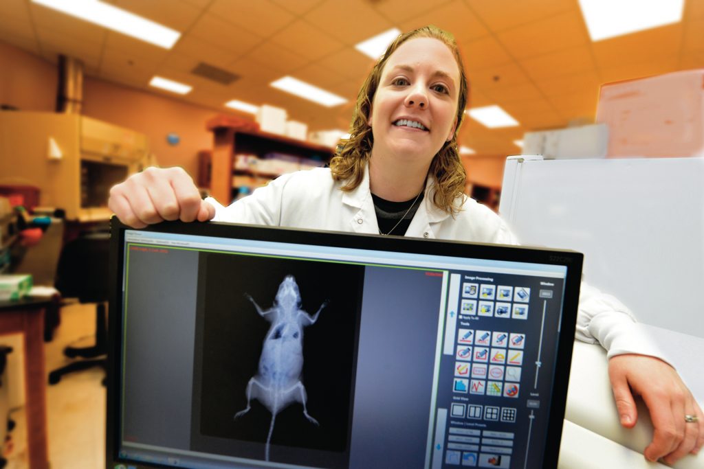
Mounting evidence of the skeleton’s endocrine role may yield new treatments for obesity.
“We know that bone is great for biomechanical motion, protecting internal organs and serving as a bank for calcium. But the skeleton has an important endocrine role in the body as well. If we can understand that role, we can come up with new avenues for treatment,” said Dr. Meghan E. McGee-Lawrence, biomedical engineer and a corresponding author of a study on the topic in the journal Molecular and Cellular Endocrinology.
When her research team deleted Hdac3, a molecule that helps regulate gene expression, from the skeletons of mice, the mice had not only weaker bones, but major metabolic changes. And although the mice continued eating normally, they lost body fat, had a low fasting-glucose level and used insulin efficiently.
“The knockout mice are making something that is good for their metabolic rate,” McGee-Lawrence said. “If we can figure out what that molecule is, that is the starting point for a new therapy.”
They suspect part of the magic may be a molecule called osteoprotegerin, which helps the liver maintain healthy insulin sensitivity. OPG is also made by osteoblasts to help decrease the amount of bone broken down in the skeleton. The drug denosumab, a newer-generation osteoporosis drug, mimics the molecule’s function in the skeleton, making it a potentially good candidate to treat obesity and its related health issues, including type 2 diabetes and cardiovascular disease.
VITAMIN D DOSAGE
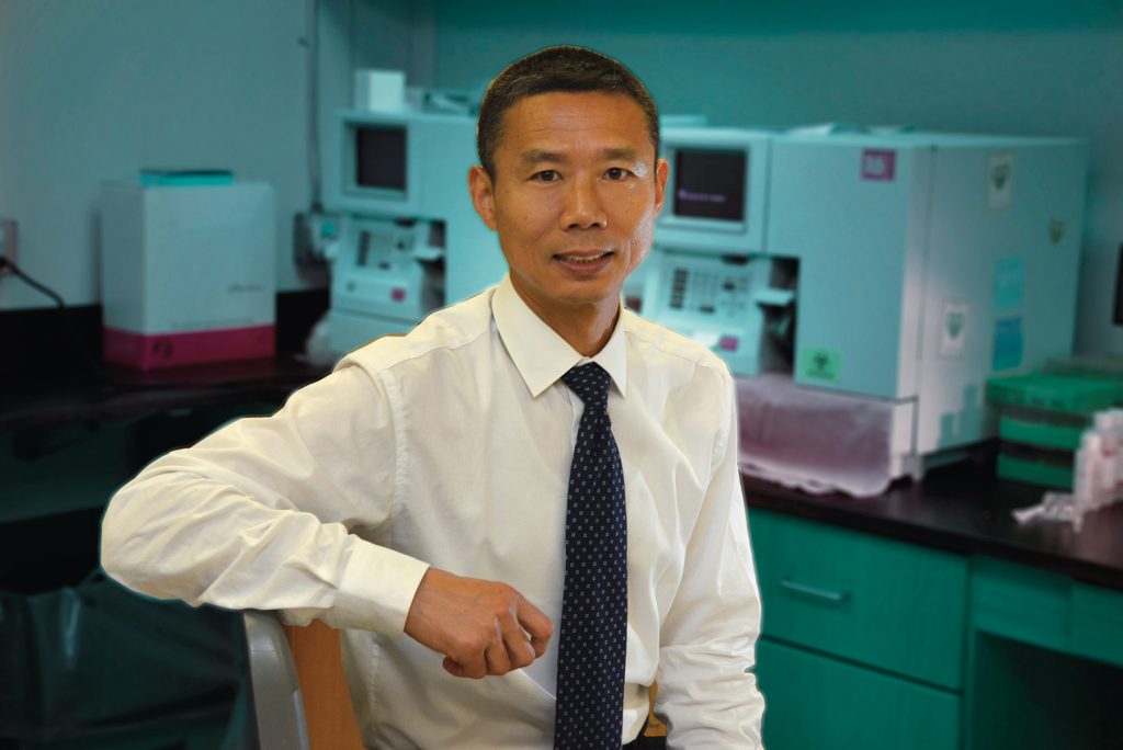
The current recommended minimum daily dose of vitamin D is not sufficient for overweight blacks, researchers report.
More than triple the dosage is needed, said Dr. Yanbin Dong, an MCG geneticist and cardiologist, noting that darker skin absorbs less sunlight and fat tends to sequester vitamin D.
The study, published in the journal BioMed Central Obesity, looked at the effects of three levels of vitamin D supplementation in 70 overweight-to-obese black adults living in the southeastern United States who appeared healthy other than a low level of vitamin D in the bloodstream.
The Institute of Medicine recommends a daily intake of 600 international units of vitamin D for most children and adults, with 4,000 IUs being the recommended maximum. But the researchers found that between 2,000 and 4,000 IUs is needed in overweight blacks to optimize blood levels of vitamin D within 16 weeks. Lower levels increase the risk for bone-weakening rickets and other problems.
The 4,000 upper-limit dose restored the healthy blood level quicker – by eight weeks – and helped suppress parathyroid hormone, which thwarts vitamin D’s ability to absorb calcium, said Dong, the study’s corresponding author.
“We hope these studies will give physicians better guidelines for some of their patients,” said Dong.
Dr. Jigar Bhagatwala, a research resident in the MCG Department of Medicine, is the study’s first author. The research was funded by the National Institutes of Health and the American Heart Association.
WAKE-UP CALL
A wristband that records motion throughout a 24-hour cycle may be an inexpensive, safe way to determine which patients with major depressive disorder will respond best to commonly prescribed drugs such as Prozac.
Selective serotonin reuptake inhibitors, or SSRIs, are today’s mainstay for depression treatment, but fine-tuning doses and prescriptions can take months, said Dr. W. Vaughn McCall, chairman of the Department of Psychiatry and Health Behavior.
His study in the Journal of Psychiatric Research indicates that the wristband may help by identifying night owls, who appear to be the best responders to SSRIs.
He hopes the finding will help refine the treatment protocol to expedite depression relief.
KIDNEY COMPENSATION
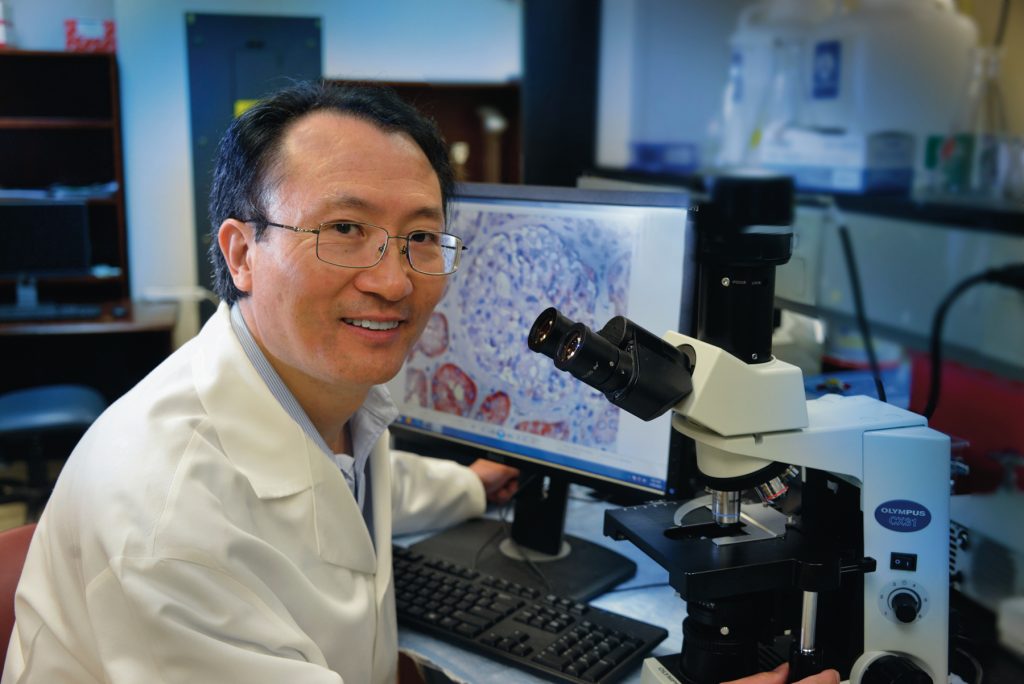
Scientists have found an explanation for the century-old observation that removing a kidney makes the remaining one grow.
Dr. Jian-Kang Chen, an MCG pathologist and co-corresponding author of a paper on the topic published in The Journal of Clinical Investigation, attributes the growth to an increase in protein-building amino acids.
A decade ago, Chen published a paper in the Journal of the American Society of Nephrology showing that activation of a protein called mTOR is a major player. However, the growth signal that activated mTOR was still unknown.
Now, the research team, which included scientists at Vanderbilt University School of Medicine, Texas A&M University and Feinberg School of Medicine at Northwestern University, has found the increased availability of amino acids prompts increased activation of mTORC1 in mice. They are now seeking to control the growth, hypothesizing that the increased size of the remaining kidney ultimately impedes kidney function.
NO OSTEOPOROSIS LINK
Postmenopausal women with kidney or bladder stones are not at increased risk for osteoporosis, but they do have about a 15 percent increased risk of another stone, physician-scientists report.
Researchers looked at data on approximately 150,000 postmenopausal women and found, despite the two conditions being clearly associated in men, the same did not hold true for women, said Dr. Laura D. Carbone, chief of the MCG Section of Rheumatology.
“We know in men that if you have a kidney stone, you are more likely to have osteoporosis,” said Carbone, corresponding author of the study in the Journal of Bone and Mineral Research. “But we found that a woman having a kidney stone is not a risk factor for osteoporosis. However, having one urinary tract stone does put women at increased risk for a second stone.”
“We wanted women and their physicians to have this information,” said Dr. Monique Bethel, a research resident in the MCG Department of Medicine and study co-author. “If the two relate and a patient who has not been screened for osteoporosis comes to the office with a kidney stone, her physician might have been concerned she also has a higher risk for osteoporosis. Our studies indicate she likely does not.”
KEY RECEPTOR
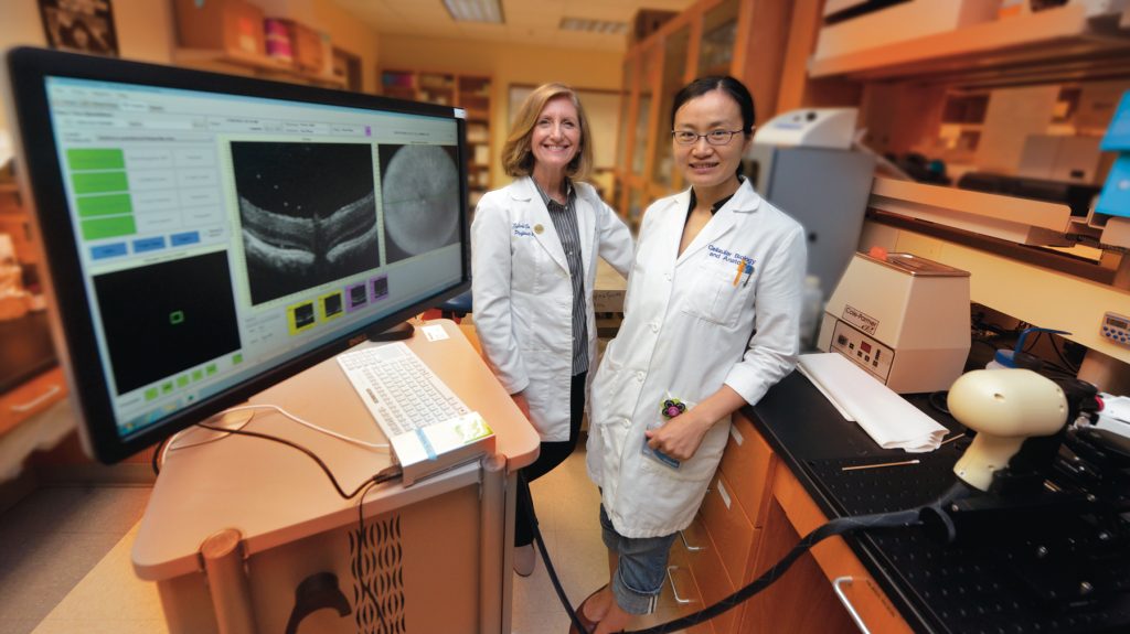
A receptor that is already a target for treating neurodegenerative disease also appears to play a key role in supporting the retina, scientists report.
Without sigma 1 receptor, the Müller cells that support the retina can’t seem to control their own levels of destructive oxidative stress and, consequently, can’t properly support the neurons that transform light into images, scientists report in the journal Free Radical Biology and Medicine.
Without support, well-organized layers of retinal cells begin to disintegrate, and vision is lost to diseases such as retinitis pigmentosa, diabetic retinopathy and glaucoma, said Dr. Sylvia Smith, retinal cell biologist and chairwoman of the MCG Department of Cellular Biology and Anatomy.
The surprising finding makes the sigma 1 receptor a treatment target for these retinal diseases, said Smith, the study’s corresponding author. It has implications as well for other diseases characterized by oxidative stress, including cardiovascular disease, cancer and neurodegenerative disease.
What most surprised the scientists was that simply removing sigma 1 receptor from Müller cells significantly increased levels of reactive oxygen species, or ROS, indicating the receptor’s direct role in the oxidative stress response, Smith said. Upon comparing the sigma 1 receptor knockouts with normal mice, they found significantly decreased expression in the knockouts of several well-known antioxidant genes and their proteins. Further examination showed a change in the usual stress response.
These genes that make natural antioxidants contain antioxidant response element, or ARE, which in the face of oxidative stress is activated by NRF2, a transcription factor that usually stays in the fluid part of the cell, or cytoplasm. NRF2 is considered one of the most important regulators of the expression of antioxidant molecules. Normally, the protein KEAP1 keeps it essentially inactive in the cytoplasm until needed, then it moves to the cell nucleus where it can help mount a defense. Deleting the sigma receptor in the Müller cells altered the healthy response: NRF2 expression decreased while KEAP1 expression increased. ROS levels increased as well.
The study is believed to provide the first evidence of the direct impact of the sigma 1 receptor on the levels of NRF2 and KEAP1, the researchers write.
The research was supported by the National Eye Institute and GRU’s James and Jean Culver Vision Discovery Institute.
Dr. Jing Wang, an MCG assistant research scientist, is the study’s first author.
PATHWAYS FOR PROTECTION
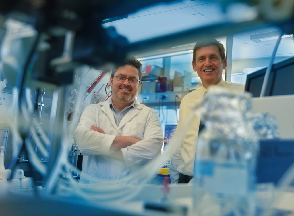
MCG researchers have found that inflamed kidney cells produce one of the same inflammation-suppressing enzymes fetuses use to survive.
The inflamed kidney cells of laboratory animals produce the enzyme indoleamine 2,3-dioxygenase, or IDO, unleashing a chain of events that can eliminate the damaged protein produced during inflammation, said Dr. Tracy L. McGaha, an MCG immunologist and corresponding author of the study featured on the cover of the Journal of Immunology. Prolonged inflammation can jeopardize the kidneys’ ability to filter blood.
The researchers also found evidence of the same protective mechanism in kidney tissue from humans with a variety of inflammation-related conditions.
“When the kidney cells couldn’t make IDO, the inflammation caused them to die,” McGaha said.
“What we are realizing is that most diseases have common pathways to either inflammation, fibrosis or recovery,” said Dr. Michael P. Madaio, an MCG nephrologist and a study co-author. “Dr. McGaha is discovering those pathways or identifying new pathways in inflammation and protection.”
Drs. David Munn and Andrew Mellor, MCG researchers, reported in 1998 that the fetus expresses IDO to avoid being targeted by the mother’s immune system. Later studies found tumors use it as well.
SEEDS OF HYPERTENSION
Chronically traumatized children typically have higher blood pressure as young adults than their peers, researchers report.
The average difference of 10 points in systolic pressure – the pressure while the heart is contracting – by early adulthood increases the risk of eventual hypertension and coronary artery disease, said Dr. Shaoyong Su, an MCG genetic epidemiologist.
“Ten points is a big difference,” said Su, corresponding author of the study in the American Heart Association journal Circulation. “You can predict that five years later, these young people may be hypertensive.”
The study looked at data collected from young people, now a mean age of 30, who are part of a long-term study at MCG’s Georgia Prevention Institute looking at cardiovascular risk factor development. About 70 percent of the children from the Richmond County public school system reported at least one adverse childhood experience; 18 percent reported more than three.
“We hope these studies will reinforce the need to screen children and young adults for adverse childhood experiences so this increased risk can be identified early to enhance resiliency and recovery and lessen the burden of cardiovascular disease later in life,” said Su, citing abuse, neglect and household dysfunction as examples of adverse childhood experiences. “First, we have to know and accept that these difficult problems occur to some extent in the majority of our children.”


