A new “image analysis pipeline” is giving scientists rapid new insight into how disease or injury have changed the body, down to the individual cell.
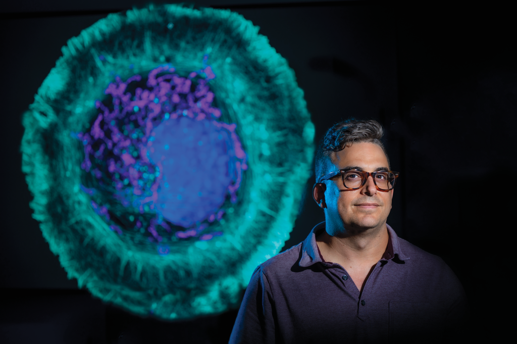
It’s called TDAExplore, which takes the detailed imaging provided by microscopy, pairs it with a hot area of mathematics called topology, which provides insight on how things are arranged, and the analytical power of artificial intelligence to give, for example, a new perspective on changes in a cell resulting from ALS and where in the cell they happen, says Dr. Eric Vitriol, MCG cell biologist and neuroscientist.
At least in the analyzing data department, computers have our brains beat, he says, not just in their objectivity but in the amount of data they can assess. Computer vision, which enables computers to pull information from digital images, is a type of machine learning that has been around for decades, so he and his colleague and fellow corresponding author Dr. Peter Bubenik, a mathematician at the University of Florida and an expert on topological data analysis, decided to partner the detail of microscopy with the science of topology and the analytical might of AI. Topology and Bubenik were key, Vitriol says.
Topology is “perfect” for image analysis because images consist of patterns, of objects arranged in space, he says, and topological data analysis (the TDA in TDAExplore) helps the computer also recognize the lay of the land, in this case where actin — a protein and essential building block of the fibers, or filaments, that help give cells shape and movement — has moved or changed density. It’s an efficient system, that instead of taking literally hundreds of images to train the computer how to recognize and classify them, it can learn on 20 to 25 images.
Part of the magic is the computer is now learning the images in pieces they call patches. Breaking microscopy images down into these pieces enables more accurate classification, less training of the computer on what “normal” looks like, and ultimately the extraction of meaningful data.
The bottom line is that the unique coupling used in TDAExplore efficiently and objectively tells the scientists where and how much the perturbed cell image differs from the training, or normal, image, information which also provides new ideas and research directions, he says.
The ability to get more and better information from images ultimately means that information generated by basic scientists like Vitriol, which often ultimately changes what is considered the facts of a disease and how it’s treated, is more accurate. That might include being able to recognize changes, like those the new system pointed out inside the cell, that have been previously overlooked.