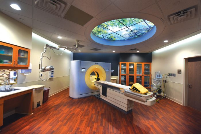
Along with magnetic resonance imaging (MRI), computed tomography (CT) was voted by physicians in 2001 as the top medical innovation of the previous 25 years, and for good reason. The CT gives physicians a quick, noninvasive look inside the body, which makes it a great way to detect a variety of diseases and conditions, and it’s very helpful when it comes to studying the effectiveness of treatments.
“This is a big part of our clinical trial work,” said Dr. James Rawson, chair of the Department of Radiology and Imaging. “There are a number of clinical trials that the Georgia Cancer Center is a part of; we support the imaging that occurs for these trials.”
For example, CT scans are integral to the university’s work with the NCI-Molecular Analysis for Therapy Choice (NCI-MATCH) clinical trial, which explores treating patients based on the molecular profiles of their tumors.
Patients with advanced solid tumors and lymphomas that are not responsive to chemotherapy or other standard therapies are screened with a tumor biopsy.
Tumors are analyzed to identify gene abnormalities that may respond to targeted drugs selected for the trial. CT scans are done periodically to track tumor responsiveness to the identified drug.
“The CT is really a workhorse for cancer imaging,” said Rawson.
A CT scan uses a computer to combine X-rays taken from different angles to produce cross-sectional images of internal organs, bones, soft tissue and blood vessels. The more images, the better the look inside, and the number of images being provided is increasing with each generation of scanner.
“When I started in radiology, a CT scan of a chest was 20 images,” Rawson said. “When I was junior faculty here, it was 200 images, and now, it can be as high as 2,000 images.”
The current CT facility, part of the groundbreaking 15-year alliance with Philips Healthcare, was planned out with patients at the design table. The result: a clean, inviting area with a calming skylight built in above the bed.


